


Archaeologists have used advanced technology on an extraordinary pair of patients: two ancient Egyptian priests who died more than 2,000 years ago.
Radiologists at Keck Medicine of USC have carried out detailed CT scans on the mummified remains of Nes-Min, who lived around 330 BCE, and Nes-Hor, who died roughly 190 BCE.
Using a state-of-the-art 320-slice scanner, the team captured hundreds of cross-sectional images while each mummy remained inside the lower half of its sarcophagus, each weighing around 200 pounds.
The scans revealed striking detail, including facial features such as eyelids and lips, allowing researchers to visualise the wrapped figures as recognisable individuals.
TRENDING
Stories
Videos
Your Say
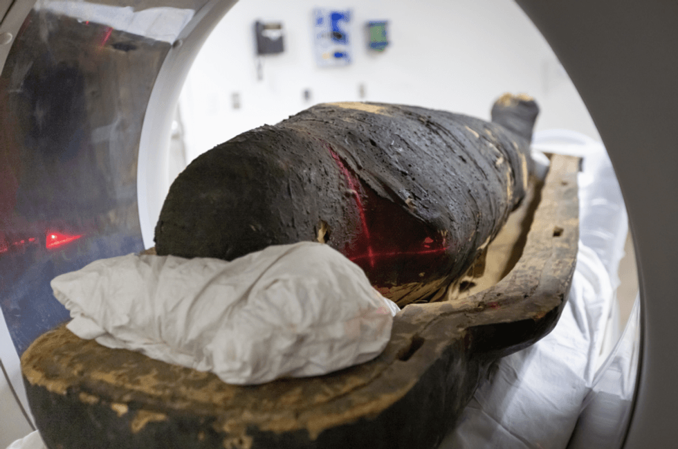
Both priests were found still clothed in linen shrouds darkened with age, with Nes-Min additionally adorned with a beaded net garment and colourful strands.
The imaging uncovered health conditions familiar to modern medicine.
Nes-Min showed a collapsed lumbar vertebra in his lower spine, consistent with long-term strain and age-related degeneration, likely causing chronic back pain.
Nes-Hor, meanwhile, suffered from extensive tooth decay and severe deterioration in one hip joint - damage typically associated with restricted mobility and significant pain.

Using a state-of-the-art 320-slice scanner, the team captured hundreds of cross-sectional images
|KECK MEDICINE OF USC/RICHARD CARRASCO III
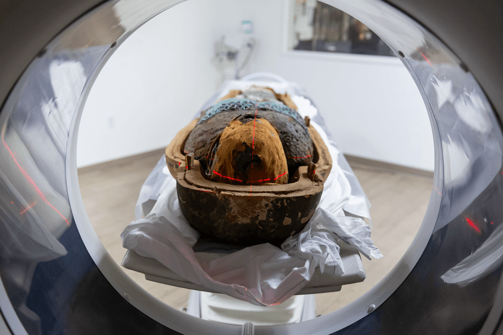
Researchers captured images while each mummy remained inside the lower half of its sarcophagus
|KECK MEDICINE OF USC/RICHARD CARRASCO III
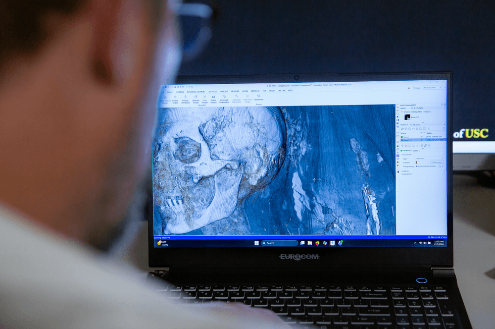
The scans revealed striking detail, including facial features such as eyelids and lips
|KECK MEDICINE OF USC/RICHARD CARRASCO III
Despite being identified as the younger of the two priests, bone analysis suggested Nes-Hor actually lived longer than Nes-Min.
“These scans provide a treasure trove of information made possible by Keck Medicine's access to the latest in high-level scanning, coupled with the team's expertise,” said Dr Summer Decker, director of the USC Center for Innovation in Medical Visualisation.
The CT scans also revealed burial objects concealed within Nes-Min’s wrappings for more than 2,000 years.
Small artefacts shaped like scarab beetles and a fish were found alongside his body and could be measured and examined digitally without disturbing the remains.
REMARKABLE DISCOVERIES - READ MORE:
The CT scans also revealed burial objects concealed within Nes-Min’s wrappings
|KECK MEDICINE OF USC/RICHARD CARRASCO III
Following the scans, Dr Decker and colleague Jonathan Ford created detailed three-dimensional digital reconstructions.
Using medical-grade 3D printers, the team produced full-scale physical replicas of both priests’ skulls, spines and hips, as well as the hidden burial objects.
“The high-resolution images have revealed things that were previously unknown and helped create a picture of what their lives were like,” Decker said.
The mummies, alongside the digital models and 3D-printed replicas, will feature in Mummies of the World: The Exhibition.
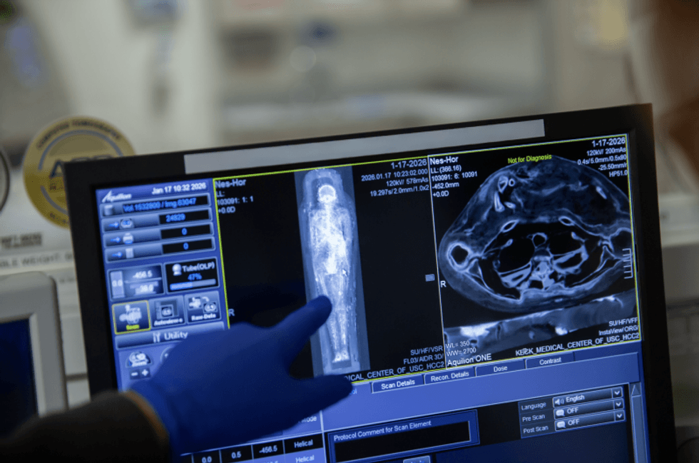
The imaging uncovered health conditions familiar to modern medicine
|KECK MEDICINE OF USC/RICHARD CARRASCO III
The showcase will commence on February 7 at the California Science Centre.
The exhibition first opened at the venue in 2010 before touring internationally - and now returns to Los Angeles as its final stop, showcasing mummies not previously displayed in the city.
“Mummies have long been a mystery. Seeing beneath the surface to reveal the specific lived experience of individuals is incredibly exciting,” said anthropologist Dr Diane Perlov.
“This modern scientific technology offers us a powerful window into the world of ancient people and past civilizations that might otherwise be lost.”
Our Standards:
The GB News Editorial Charter